Scanning Helium Microscopy (SHeM)
Home > Equipment > Testing and validation > Scanning Helium Microscopy (SHeM)
Scanning Helium Microscopy (SHeM)

Developed by the University of Newcastle in collaboration with researchers from Cambridge University, the Newcastle Scanning Helium Microscope (SHeM) is an extremely surface-sensitive imaging technique. The tool, one of only three in the world, probes samples using neutral helium atoms instead of the more traditional light or electrons. Advantages include minimal sample preparation requirements (no charge reducing coatings required), a complete lack of sample damage as can occur under beams of electrons or laser light, and no penetration of the probe into the surface at all (extreme sensitivity to thin, transparent films).
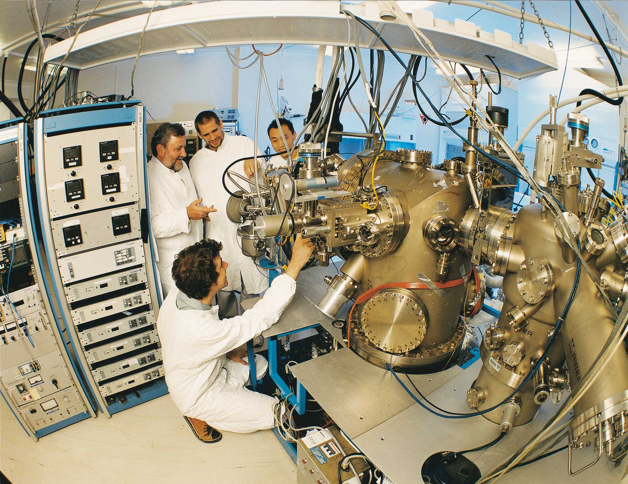
List of available equipment
TOOL MAKE AND MODEL
KEY DIFFERENTIATOR
LOCATION
Scanning Helium Microscope (SHeM)- Custom
Scanning Helium Microscope (SHeM)
Materials Node University of Newcastle
Description
A custom built highly surface-sensitive imaging system
Related Information
Maximum sample size ~ 16 x 16 mm, resolution ~ 1 micron
Tool Contact
anff@newcastle.edu.au